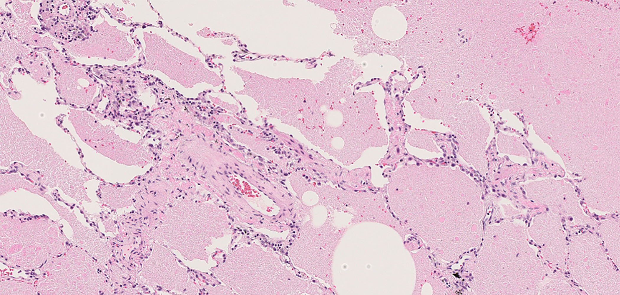
How do doctors diagnose pulmonary alveolar proteinosis?
Ninety-two percent of pulmonary alveolar proteinosis is autoimmune alveolar proteinosis caused by anti-GM-CSF autoantibodies. The order of probability is autoimmune pulmonary alveolar proteinosis → secondary pulmonary alveolar proteinosis → hereditary pulmonary alveolar proteinosis, and so doctors explore possibilities in this order.
Main examinations
- 1. CT scanHigh-resolution CT scan of the lungs
- 2. Blood testMeasurement of Serum anti-GM-CSF autoantibody concentration
- 3. BronchoscopyConfirmation of surfactant in the alveoli
In the case of autoimmune alveolar proteinosis:
1) If a high-resolution CT scan of the lungs shows ground-glass opacities (GGO), pulmonary alveolar proteinosis is suspected.

2)The anti-GM-CSF autoantibody concentration is measured with a blood test; a reading of 1.7 U/ml or higher is considered diagnostic. The stick type immunochromatography assay (Line Check APAP, Nobelpharma, Co., Ltd.) is also available. If the test is positive, there is a high possibility of autoimmune pulmonary alveolar proteinosis.
3)To confirm the diagnosis, it is usual to check for a milk-like appearance of bronchoalveolar lavage fluid.
 Photo : Saline solution being pumped into the bronchi using a bronchoscope
Diagnoses will be made in the order of 1) → 2) → 3, but please consult with your doctor carefully as he or she may have his or her own opinion.
In the case of secondary pulmonary alveolar proteinosis:
Tests 1), 2), and 3)are required, but test 2)is usually negative. In addition, for diagnosis, it is necessary to have an underlying disease that is reported as the cause of pulmonary alveolar proteinosis. Reported diseases include myelodysplastic syndromes, leukemia, infections, and collagen diseases.
In the case of hereditary pulmonary alveolar proteinosis:
Tests 1), 2), and 3)are still required, but if 2)is negative and there is no known underlying disease that would cause secondary alveolar proteinosis, hereditary alveolar proteinosis is suspected. It usually takes many months to locate and determine the gene responsible.

