Characteristics of chest CT findings of pulmonary alveolar proteinosis
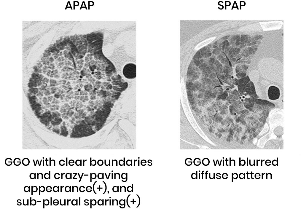
The chest CT images of autoimmune pulmonary alveolar proteinosis and secondary pulmonary alveolar proteinosis are both characterized by GGO (Ground-glass opacities). In autoimmune pulmonary alveolar proteinosis, the spread of GGO is indicated by a patchy pattern with clear borders as shown in the figure below:
A “crazy-paving” appearance (71%) that looks like cantaloupe skin and subpleural GGO that do not reach the subpleural area sparing (71%) are noticeable. By contrast, the above findings are less typical in secondary pulmonary alveolar proteinosis, at 14% and 33%, respectively. Moreover, the distribution of GGO spreads predominantly to the lower lung fields in autoimmune pulmonary alveolar proteinosis, but not in secondary alveolar proteinosis. GGO are also the main finding in chest CT of hereditary alveolar proteinosis, but since it is rare, it is not clear whether it has characteristics similar to those of the above two diseases.

Autoimmune alveolar proteinosis (axial view)

Secondary alveolar proteinosis (axial view)


